Doç. Dr. Serdar ÖZATEŞ
Assoc. Prof. Dr. Serdar ÖZATES
Göz Hastalıkları Uzmanı
Ophthalmology Specialist
×
Assoc. Prof. Dr. Serdar ÖZATEŞ
Eyelid Diseases and Oculoplastic Surgery
All surgical interventions on the eyelids, tear ducts and structures around the eye comprise oculoplastic surgery. Oculoplastic surgeries can be performed for aesthetic and cosmetic purposes as well as to correct disorders or diseases around the eye.
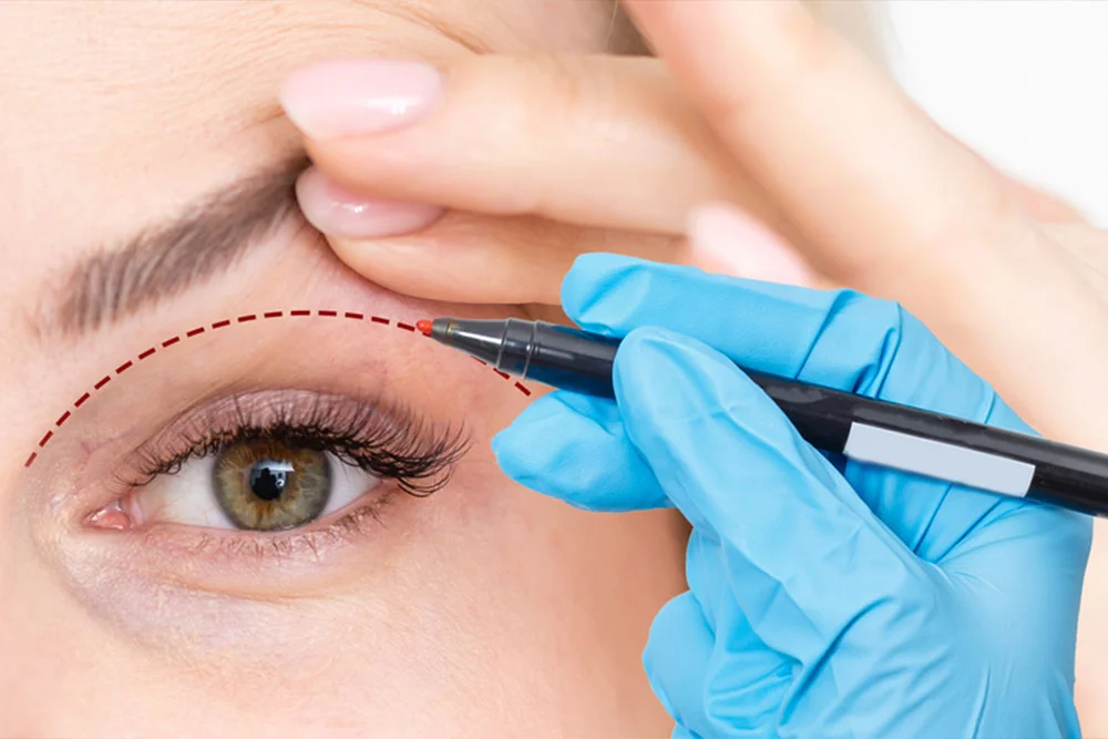
Blepharoplasty
The condition referred to as blepharochalasis is the sagging of the upper or lower eyelid skin excess and subcutaneous fat tissue hernia. Upper or lower eyelid blepharoplasty is applied in its treatment. Depending on the patient’s needs and wishes, the surgery is planned specifically for each patient. The aim of the blepharoplasty procedure is to remove excess skin and fat tissue in accordance with the patient’s wishes and age without impairing the function of the eyelid.
Ptosis / Droopy Eyelid
Ptosis can be described as the upper eyelid droop and not being able to open fully. In patients with ptosis, there is an anatomical or functional problem in the muscle that lifts the eyelid. The exact cause of eyelid drooping should be determined. Treatment varies depending on the cause of eyelid drooping. As a surgical treatment, the levator resection technique (shortening the muscle that lifts the upper eyelid) or the frontal suspension technique (attaching the eyelid to the forehead with stitches) can be applied.
Entropion / Eyelid Turning Inward
Entropion is the turning of eyelid inwards. Entropion causes eyelash to get in contact with the ocular surface. Due to constant contact, recurrent eye surface infections and decreased corneal transparency may cause decreased vision quality. First, the cause of entropion should be determined and the treatment method is decided.
Ectropion / Eyelid Turning Outward
Ectropion is the turning of eyelid outwards. Outwards turning of eyelid edge leaves the eye surface exposed. Prolonged ocular surface exposure may cause dry eyes, recurrent infections and decreased corneal transparency, which may cause decreased vision quality. First, the cause of ectropion should be determined and the treatment decision is made.
Eyelid Tumors
Many types of tumors, benign or malignant, can develop on and around the eyelid. Lesions that grow and spread rapidly to surrounding tissue, show color change, and have a wound appearance that does not heal are considered suspicious for tumors. The treatment of tumors is surgical, and the surgical plan varies according to the location, type, and size of the tumor tissue.
Eyelid Surgery Process
Before Surgery
Surgery Day
After Surgery
Lacrimal Duct Obstruction
Tear secretion drains into the nose through channels located on the medial edge of the eyelids. Lacrimal ducts can be blocked congenitally or due to acquired reasons such as recurrent infections.
Congenital Lacrimal Duct Obstruction
The development of the lacrimal duct sometimes lags behind and is closed at birth and opens during delivery or after birth. The first symptoms are watery eyes, epiphora and recurrent infections that resistant to treatment. Since the possibility of the lacrimal duct opening on its own is high in the first year of life, only massaging the lacrimal system is recommended in treatment. If the blockage does not open with massage by the end of the age of 1, the lacrimal duct is opened with a procedure called nasolacrimal probing. In this procedure, only the lacrimal duct is intervened. The procedure is performed with short-term general anesthesia.
Before Surgery
Day of Surgery
After Surgery
Acquired Tear Duct Obstruction
Tear duct obstruction may develop due to recurrent infections or advancing age. The first symptoms are epiphora in a closed environment and during rest, and recurrent infections that resistant to treatment. It is confirmed with examination that the tear duct is blocked. The treatment for tear duct obstruction in adults is surgery. In the surgery called dacryocystorhinostomy, a silicone tube is placed between the tear ducts and the nasal cavity to provide new tear duct passage. When the healing process is complete, the placed silicone tube is removed approximately 6 months after the surgery.
Before Surgery
Surgery Day
After Surgery
You can create your appointment from Ophthalmology Specialist Assoc. Prof. Dr. Serdar ÖZATEŞ clinic by filling out the form below.